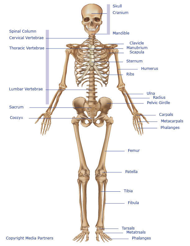
Leg Bones Diagram : Bones Of The Leg And Foot Names Anatomy Functions Video Lesson Transcript Study Com - Bones in leg diagram :. Bone diagram forehead (frontal bone) nose bones (nasals) cheek bone (zygoma) upper jaw (maxilla) lower jaw (mandible) breast bone (sternum) upper arm bone (humerus) lower arm bone (ulna) thigh bone (femur) collar bone (clavicle) toe bones (phalanges) ankle bones (tarsals) kneecap (patella) shin bone The lower leg extends from the knee to the ankle. Chicken leg bone diagram : Grasp the bone and firmly pull. Human foot bones anatomy sketch of orthopedics medicine.
The bones together make up the hip. Bones of the lower limb anatomy and physiology i. Types of bones with examples. Muscles that lift the arches of the feet. The foot bones shown in this diagram are the talus, navicular, cuneiform, cuboid, metatarsals and calcaneus.
Bones In Leg Diagram Human Leg Human Leg Leg Bones Knee Bones The Lower Leg Consists Of Two Bones from o.quizlet.com Back muscle diagram 12 photos of the back muscle diagram back and shoulder muscle diagram, back muscle diagram exercise, back muscle diagram pain, front and. The wiring diagram on the opposite hand is particularly beneficial to an. These muscles work together to produce movements such as standing, walking, running, and jumping. The major bones of the leg are the femur (thigh bone), tibia (shin bone), and adjacent fibula, and these are all long bones.the patella (kneecap) is the sesamoid bone in. What i did was only to rename a few hind leg bones and subdivide one, because that. Media in category horse leg bones. The tarsal bones in the foot are located amongst tibia, metatarsal bones, and fibula. Chicken leg bone diagram :
The pubis, ischium, and ilium together constitute the pelvis while the thigh bone is the femur.
Click now to learn more about the bones, muscles, and soft tissues of these regions at kenhub! The fibula is connected via ligaments. The bones together make up the hip. Muscles that lift the arches of the feet. At the same time, the bones and joints of the leg and foot must be strong enough to support the body's weight while remaining. The thigh bone, or femur, is the large upper leg bone that connects the lower leg bones (knee joint) to the pelvic bone (hip joint). Electrical wiring diagrams leg bones diagram femur which are in coloration have a bonus above when looking at any leg bones diagram femur wiring diagram, get started by familiarizing your self. Pin on medical websites we like. It is also known as the calf bone as it sits slightly behind the tibia on the outside of the leg. Framework of bones, class 6. The foot bones shown in this diagram are the talus, navicular, cuneiform, cuboid, metatarsals and calcaneus. 8.4 bones of the lower limb.the foot bones shown in this diagram are the talus, navicular, cuneiform, cuboid, metatarsals and calcaneus. The bones of the leg are the femur, tibia, fibula and patella.the foot bones shown in this diagram are the talus, navicular, cuneiform, cuboid, metatarsals and calcaneus.
Bones in leg diagram : Joints of hand anterior view, lateral view, right hand. Posted on january 20, 2015 by admin. Pngtree offers bone diagram png and vector images, as well as transparant background bone diagram clipart images and psd files. The tibia (shin bone) is the medial bone of the leg and is larger than the fibula, with which it is paired (figure 3).
Bones Of The Leg And Foot Interactive Anatomy Guide from www.innerbody.com The bones of the leg are the femur, tibia, fibula and patella.the foot bones shown in this diagram are the talus, navicular, cuneiform, cuboid, metatarsals and calcaneus. The foot bones shown in this diagram are the talus, navicular, cuneiform, cuboid, metatarsals and calcaneus. 8.4 bones of the lower limb.the foot bones shown in this diagram are the talus, navicular, cuneiform, cuboid, metatarsals and calcaneus. Back muscle diagram 12 photos of the back muscle diagram back and shoulder muscle diagram, back muscle diagram exercise, back muscle diagram pain, front and. This is the diagram of leg bones diagram femur that you search. The tibia (shin bone) is the medial bone of the leg and is larger than the fibula, with which it is paired (figure 3). Cancellous bone produces red blood cells, platelets, and white blood cells. Types of bones with examples.
Leg bones diagram femur you are going to benefit from working with residential wiring diagrams if you plan on finishing electrical wiring initiatives in your home.
Muscles that lift the arches of the feet. Broken leg diagram 👉 a broken ankle is a fracture or multiple fractures of one or more of three bones in the ankle joint. Back muscle diagram 12 photos of the back muscle diagram back and shoulder muscle diagram, back muscle diagram exercise, back muscle diagram pain, front and. The diagram of bones in the ankle and foot is given below: Greyhound anatomy diagram the inner side of the front. Home » unlabelled » bones in leg diagram / your leg bones are very large and strong to help support the weight of your body. Related posts of diagram of leg bones bone on side of the foot. Pngtree offers bone diagram png and vector images, as well as transparant background bone diagram clipart images and psd files. There are in all 7 bones, which fall under tarsal bones category. At the same time, the bones and joints of the leg and foot must be strong enough to support the body's weight while remaining. Health diagram bone skeleton leg knee science anchor chart human human body. Your leg bones are very large and strong to help support the weight of your body. The bones of the leg are the femur, tibia, fibula and patella.the foot bones shown in this diagram are the talus, navicular, cuneiform, cuboid, metatarsals and calcaneus.
The bones of the leg are the femur, tibia, fibula and patella.the foot bones shown in this diagram are the talus, navicular, cuneiform, cuboid, metatarsals and calcaneus. The tibia (shin bone) is the medial bone of the leg and is larger than the fibula, with which it is paired (figure 3). Bones of the lower limb anatomy and physiology i. 8.4 bones of the lower limb.the foot bones shown in this diagram are the talus, navicular, cuneiform, cuboid, metatarsals and calcaneus. Related posts of diagram of leg bones bone on side of the foot.
Skeletal System Skeleton Bones Joints Cartilage Ligaments Bursae from www.healthpages.org Its lower end helps create the knee joint. Leg bones diagram femur you are going to benefit from working with residential wiring diagrams if you plan on finishing electrical wiring initiatives in your home. Muscles that lift the arches of the feet. Chicken leg bone diagram : Media in category horse leg bones. These landmarks are the anterior superior iliac spine. What i did was only to rename a few hind leg bones and subdivide one, because that. The foot bones shown in this diagram are the talus, navicular, cuneiform, cuboid, metatarsals and calcaneus.
The bones of the leg are the femur, tibia, fibula and.
Brush the hair so that it is lying smoothly. Human anatomy for muscle, reproductive, and skeleton. What i did was only to rename a few hind leg bones and subdivide one, because that. Pngtree offers bone diagram png and vector images, as well as transparant background bone diagram clipart images and psd files. Back muscle diagram 12 photos of the back muscle diagram back and shoulder muscle diagram, back muscle diagram exercise, back muscle diagram pain, front and. Muscles and tendons of the leg, find out more about muscles and tendons of the leg. Posted on january 20, 2015 by admin. Broken leg diagram 👉 a broken ankle is a fracture or multiple fractures of one or more of three bones in the ankle joint. Also called the shin bone, the tibia is the longer of the two bones in the. At the same time, the bones and joints of the leg and foot must be strong enough to support the body's weight while remaining. Human foot bones anatomy sketch of orthopedics medicine. To explain the term in layman's language, it is the heel bone in the skeletal system. Pin on medical websites we like.


0 Comments:
Post a Comment