Ct Pelvis Anatomy Muscles : Radiology Images / Key facts about the muscles of the pelvic floor.. Key facts about the muscles of the pelvic floor. A commonly conducted imaging test on the abdomen and pelvis region is the abdominopelvic ct(10). It is bounded on either side by the ilium; The muscles of the pelvis, hip and buttock anatomical chart shows how each muscle in this area of the body works with the others, and the various minor systems within the major ones. N patient preparation n patient position n scanogram.
Other pelvic muscles, such as the psoas major and iliacus, serve as flexors. Pelvic anatomy mri variant anatomy pelvic viscera. Their main function is contractibility. The tensor fascia lata and sartorious muscles originate from the anterior superior iliac spine. This mri male pelvis axial cross sectional anatomy tool is absolutely free to use.
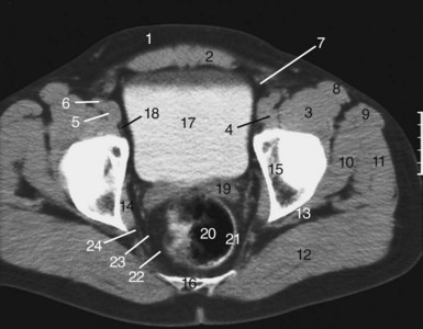
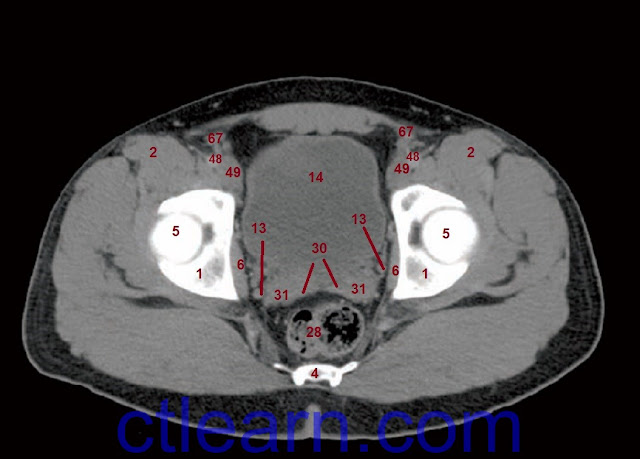
Pelvic Floor Muscles Anatomy Ct | Review Home Co from radiologykey.com 3 enumerate the muscles of true pelvis. Abdominal and pelvic anatomy encompasses the anatomy of all structures of the abdominal and pelvic cavities. Use the mouse scroll wheel to move the images up and down alternatively use the tiny arrows (>>) on both side of the image to move the images. This mri male pelvis axial cross sectional anatomy tool is absolutely free to use. Clinical anatomy of the pelvis. The main functions of the neck muscles are to permit movements of the neck or head and to provide structural support of the head. Hint you are sitting on it right now. The greater or false pelvis (pelvis major).—the greater pelvis is the expanded portion of the cavity situated above and in front of the pelvic brim.
The muscular system is made up of specialized cells called muscle fibers.
A commonly conducted imaging test on the abdomen and pelvis region is the abdominopelvic ct(10). These muscles, including the gluteus maximus and the hamstrings, extend the thigh at the hip in support of the body's weight and propulsion. Learn about anatomy muscles pelvis with free interactive flashcards. The greater or false pelvis (pelvis major).—the greater pelvis is the expanded portion of the cavity situated above and in front of the pelvic brim. Their main function is contractibility. 13 what portion of the bony pelvis is the arrow pointing to? ƒ organs and structures of the female pelvis. 3 enumerate the muscles of true pelvis. The video covers the most. Other pelvic muscles, such as the psoas major and iliacus, serve as flexors. N patient preparation n patient position n scanogram. These 4 bones are connected by 4 joints and lined by 4 muscles. Ischial tuberosity which flexor of the knee attaches here?
3 enumerate the muscles of true pelvis. Learn about anatomy muscles pelvis with free interactive flashcards. Three knee extensors originate from the pelvis. Use the mouse scroll wheel to move the images up and down alternatively use the tiny arrows (>>) on both side of the image to move the images. This anatomy section promotes the use of the terminologia anatomica, the international standard of anatomical nomenclature.
Pelvic Muscles Anatomy Ct from i.pinimg.com Muscles, connected to bones or internal organs and blood vessels, are in charge for movement. N patient preparation n patient position n scanogram. These 4 bones are connected by 4 joints and lined by 4 muscles. 13 what portion of the bony pelvis is the arrow pointing to? Ischial tuberosity which flexor of the knee attaches here? 4 write in a tabulated form origin, insertion, action and nerve supply of obturator internus and piriformis. Evan oto female hip, muscle anatomy ss284551 ve5093. Included within the chart are gorgeous illustrations of the pelvic diaphragm, sphincter muscles, gluteus maximus.
If you want to learn how to read ct scans of the abdomen and pelvis proficiently, this video is an excellent starting point.
Evan oto female hip, muscle anatomy ss284551 ve5093. Clinical anatomy of the pelvis. The pelvis is a basin shaped bony structure formed by the combination of two pelvic bones (hip bones or innominate bones) and the sacrum. These muscles, including the gluteus maximus and the hamstrings, extend the thigh at the hip in support of the body's weight and propulsion. • describe the bony anatomy of the pelvic floor • describe the skeletal muscle of the pelvic floor • discuss the arterial supply to pelvis. It is bounded on either side by the ilium; N patient preparation n patient position n scanogram. Several additional pelvic and hip muscles are better introduced as part of a lower extremity lab, but since they are so well seen here we will look at them. Programme riczo reviews the anatomy of the pelvic girdle with a focus on the bones, joints, ligaments and muscles of the pelvis and surrounding areas. Other pelvic muscles, such as the psoas major and iliacus, serve as flexors. They support the pelvic organs especially during increases in intra abdominal pressure and also aid in urinary and faecal. A commonly conducted imaging test on the abdomen and pelvis region is the abdominopelvic ct(10). Tutorial video 'urine and bowel continence'.
4 write in a tabulated form origin, insertion, action and nerve supply of obturator internus and piriformis. This mri male pelvis axial cross sectional anatomy tool is absolutely free to use. Key facts about the muscles of the pelvic floor. If you want to learn how to read ct scans of the abdomen and pelvis proficiently, this video is an excellent starting point. • describe the bony anatomy of the pelvic floor • describe the skeletal muscle of the pelvic floor • discuss the arterial supply to pelvis.
Learn CT Scan: Anatomy CT Axial Abdomen and Pelvis Male from 4.bp.blogspot.com These 4 bones are connected by 4 joints and lined by 4 muscles. Three knee extensors originate from the pelvis. Muscles of the pelvis that cross the lumbosacral joint to attach onto the trunk were described in the previous blog post note: 0835 lotze anatomy of the pelvic floor. ƒ organs and structures of the female pelvis. Included within the chart are gorgeous illustrations of the pelvic diaphragm, sphincter muscles, gluteus maximus. Muscles, connected to bones or internal organs and blood vessels, are in charge for movement. Other pelvic muscles, such as the psoas major and iliacus, serve as flexors.
Attached to the pelvis are muscles of the buttocks, the lower back, and the thighs.
Almost every movement in the body is the outcome of muscle contraction. The pelvis (plural pelves or pelvises) is either the lower part of the trunk of the human body between the abdomen and the thighs (sometimes also called pelvic region of the trunk) or the skeleton embedded in it (sometimes also called bony pelvis, or pelvic skeleton). Muscles of the pelvis that cross the lumbosacral joint to attach onto the trunk were described in the previous blog post note: These 4 bones are connected by 4 joints and lined by 4 muscles. A commonly conducted imaging test on the abdomen and pelvis region is the abdominopelvic ct(10). • describe the bony anatomy of the pelvic floor • describe the skeletal muscle of the pelvic floor • discuss the arterial supply to pelvis. Clinical anatomy of the pelvis. A sling of muscles closes the floor of the pelvis. Programme riczo reviews the anatomy of the pelvic girdle with a focus on the bones, joints, ligaments and muscles of the pelvis and surrounding areas. This is the iliopubic line which outlines the anatomic anterior column this is the ilioischial line which outlines the anatomic posterior column. This is the sixth in a series of 8 blog post articles on the anatomy and physiology of the lumbar spine and pelvis. The pelvis is a basin shaped bony structure formed by the combination of two pelvic bones (hip bones or innominate bones) and the sacrum. Although ultrasound is frequently indicated for the primary.
Muscles, connected to bones or internal organs and blood vessels, are in charge for movement anatomy muscles pelvis. The floor is composed of two muscular layers, the levator ani/coccygeus complex and.

0 Comments:
Post a Comment